Assist. Prof. Dr . Hanan Zweer
College Of Education For Pure Sciences / Department Of Biology
First discovered in 1899, Naegleria Fowleri is a world-scale common, widespread, brain-eating amoeba. This amoeba is commonly found in freshwaters, including rivers, lakes, ponds, and hot springs, and it never lives in the oceans. It is a bacteria-feeding unicellular amoeba that belongs to the phylum Percolozoa. Naegleria Fowleri can freely live in water, soil, or in a host.

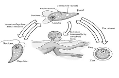
Naegleria Fowleri-caused infections may occur among children and adults while swimming and diving into contaminated waters. The infectious trophozoite passes through running water, moving into the nose. The trophozoite then sticks to the mucous membrane, moves along the olfactory nerve, and finally reaches the central nervous system’s olfactory cells. Naegleria Fowleri is the cause of primary amoebic meningoencephalitis, a highly-considered brain-damaging disease, which leads to the inflammation of the brain and membrane. As this amoeba feeds on the brain tissues, bleeding and decay develop; therefore, the patient’s death takes place in 7 to10 days.
The symptoms of primary amoebic meningoencephalitis, once developing within 2-8 days of infection, may initially appear mild but rapidly worsen. The patient may develop symptoms, which are headache, fever, chills, Photophobia, confusion, seizures, comas, irritation, vomiting, diplopia or bizarre behaviour, myocardial necrosis and abnormalities of the Cardiac rhythm, and abnormalities in some cases. Persistent symptoms may as well lead to smell and taste changes eventually. As symptoms are constantly developing, the infection concentrates in the brain base, brain stem, and cerebellum parts. This case is also manifested by a grave infection, where the most affected are olfactory mucous membranes. Severe necrotizing meningitis has also been associated with mild purulent discharge caused by this amoeba.

The Naegleria Fowleri infection, the main cause of primary amoebic meningoencephalitis, has recently increased due to thermal pollution and other environmental changes. It was proven scientifically to be attracted to heat; this amoeba can withstand surrounding temperatures at 45° C, where it boosts reproduction in summer, where the optimal temperature of an infectious trophozoite ranges from 35 to 46° C. However, if temperatures drop to 27° C and 37° C, the infectious amoeba phase can turn into a cyst and thereby be able to live at low temperatures..
Life Cycle
This infectious amoeba does not spread from one person to another by drinking water but spreads only by the nose. There are three forms of this amoeba: Cyst, Trophozoite, and Flagellate. An amoeba’s life cycle, thus, proceeds into three phases. The developing form feeds on bacteria and multiplies by binary fission. If water ionization changes or nutrients decrease, the amoeba turns into the Flagellate phase, and once surrounding conditions improve, the amoeba returns to the Trophozoite phase. As the amoeba development is permanently subject to the surrounding conditions, the amoeba turns into a cyst should these conditions be unavailable. In primary amoebic meningoencephalitis, an amoeboid trophozoite is present in cerebral myeloid fluids and tissues. In some cases, the Flagellate phase can also be found in cerebral spinal fluid.
Diagnosis
There are several methods to diagnose this amoeba;
1. Laboratory diagnosis.
2. Direct wet-mount microscopy.
3. Molecular diagnosis.
4. The examination of a stained cerebrospinal fluid smear.
Prevention
To avoid this infectious, deadly amoeba-caused disease, one must not nasally inhale fresh water. Additionally, water sports, activities, or any other diving-related acts must be avoided. Thermally polluted waters must also be avoided. Untreated waters must not be used for irrigation or nose inhalation. Pond waters should be treated with chlorine to eliminate this infectious amoeba.
References
1-Bin-Asif H. and Abid Ali S. (2018).Primary Meningoencephalittis Caused by Naegleria fowleri I: A MINI REVIEW. Baqai J. Health Sci.21( 1),
2-De Jonckheere JF. (2011). Origin and evolution of the worldwide distributed pathogenic amoebaflagellate Naegleria fowleri. Infect Genet Evol 11:1520 –1528.
3-El‐Maaty D. and Hamza RS. (2012). Primary amoebic meningoencephalitis caused by Naegleria fowleri. Pakistan University Journal, 5: 93 ‐ 104.
4-Fowler M. and Carter R.F. (1965). Acute pyogenic meningoencephalitis probably due to Acanthamnoeba spp.: A preliminary report. Brazil Medical Journal, 3: 740 – 742.
5-Grace E., ; Asbill S. and Virga K. (2015). Naegleria fowleri: Pathogenesis, Diagnosis, and Treatment Options. Antimicrobial Agents and Chemotherapy ASM.Journal.59(11): 81-6677.
6-Martínez AJ. (1985). Free-living amoebas: natural history, prevention, diagnosis, pathology, and treatment of disease. CRC Press, Boca Raton, Pp166.
7-Marciano – Cabral F.; MacLean RC.; Mensah AH. and La Pat L. (2003). Identification of Naegleria fowleri in domestic water sources by nested PCR. Applied Environmental Microbiology, 69: 5864 – 5869
8-Siddiqui R., Ali I.; Cope JR. and Khan NA. (2016). Biology and
pathogenesis of Naegleria fowleri. Acta Tropica, 164: 375 ‐ 394
59 (11), 6677-6681.
9-Shakoor S.; Beg MA.; Mahmood SF.; Bandea R.; Sriram R.and Noman F.( 2011)Primary amebic meningoencephalitis caused by Naegleria fowleri, Karachi, Pakistan. Emerg Infect:17; 61-258.
10-Visvesvara GS; Moura H; and Schuster FL. (2007). Pathogenic and opportunistic free-living amoebae: Acanthamoeba spp., Balamuthia mandrillaris, Naegleria fowleri, and Sappinia diploidea. FEMS Immunol Med Microbiol 50:1–26.
11-Yoder JS.; Straif-Bourgeois S.;Roy SL.;Moore TA.; Visvesvara GS.;Ratard RC.; Hill V.; Wilson JD.;Linscott AJ.; Crager R.; Kozak NA.; Sriram R.;Narayanan J.; Mull B.; Kahler AM.;Schneeberger C.; da Silva AJ.and Beach MJ. (2012).Deaths from Naegleria fowleri associated with sinus irrigation with tap water: a review of the changing epidemiology of primary amebic meningoencephalitis. Clin Infect.;1-7.


