

Hadeel khalaf Mohammed Alboaklah
Pharmacy
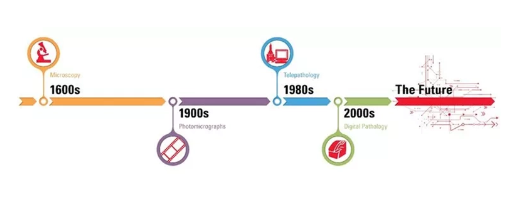
What are Digital slides are formed when slides are imagined with a scanning device to obtain a high-resolution digital image that can be observed on the screen of a computer or any device.
This process can occur following steps:
⦁ Employing high-data, automated digital pathology scanners will likely capture whole glass slides, either in bright fields or fluorescent circumstances, at a magnification analogous to a microscope.
⦁ Scanned data from digital slides can be shared through networks via particular digital pathology software.
⦁ Computerized image examination tools can also be useful to contribute to the interpretation and measurement of biomarker expression in tissue slices.
Digital Pathology?
Digital pathology unites the achievement, management, distribution, and explanation of pathology information, counting slides, and data in a digital situation.

What are the advantages of Digital Pathology?
Digital pathology will improve value in expressive ways:
⦁ Analysis procedures improved because Algorithms are objective, precise, faster than microscopy, and have swift access to previous cases. Furthermore, data Storage tolerates long-term prognostic analytics.
⦁ Diminished Errors: Rejects breakage, and barcoding lessens the risk of misidentification.
⦁ Healthier Views, Proposals live zoomed, and numerous-angle assessments afford a control panel view of figures and explanations.
⦁ Reduced Reversal Times by quicker admission to archived digital slides, reduces time-saving, data corresponding, and organizing. Rapidity admittance to samples and improves shift time versus manual analyses, particularly in multifaceted cases.
⦁ More Novelty: Large data consent pathologists converted more specialized, tolerate practices to range toward broader geographies and deliver improved tools for education and training. Professors can interpret important sections of interest accurately at the cellular and sub-cellular level, which cannot be completed readily with slides (Figure 1).
⦁ With dedicated digital pathology, educational software applications, queries, and seminars can be entrenched into the digital slides, offering background information and straight links to tissue or cellular topographies referenced in the inquiries.

Figure 1: Digital slides are frequently used by teachers for histology training. Applying digital slides and software applications is supposable to interpret substantial areas of interest.
Reference
⦁ Tizhoosh, H.R. and Pantanowitz, L., 2018. Artificial intelligence and digital pathology: challenges and opportunities. Journal of pathology informatics, 9(1), p.38.
⦁ Farahani, N., 2015. Whole slide imaging in pathology: advantages, limitations, and emerging perspectives.(2015).
⦁ Shaban, M.T., Baur, C., Navab, N. and Albarqouni, S., 2019, April. Staingan: Stain style transfer for digital histological images. In 2019 Ieee 16th international symposium on biomedical imaging (Isbi 2019) (pp. 953-956). IEEE.
⦁ Niazi, M.K.K., Lin, Y., Liu, F., Ashok, A., Marcellin, M.W., Tozbikian, G., Gurcan, M.N. and Bilgin, A., 2019. Pathological image compression for big data image analysis: Application to hotspot detection in breast cancer. Artificial intelligence in medicine, 95, pp.82-87.
⦁ Louis, D.N., Gerber, G.K., Baron, J.M., Bry, L., Dighe, A.S., Getz, G., Higgins, J.M., Kuo, F.C., Lane, W.J., Michaelson, J.S. and Le, L.P., 2014. Computational pathology: an emerging definition. Archives of pathology & laboratory medicine, 138(9), pp.1133-1138.